

Activity 5.1: Cross-section of a Leaf
Activity 5.1: Cross-section of a Leaf
Objective:
To observe the internal structure of a dicot leaf under a microscope and identify the tissues involved in photosynthesis.
Materials Required:
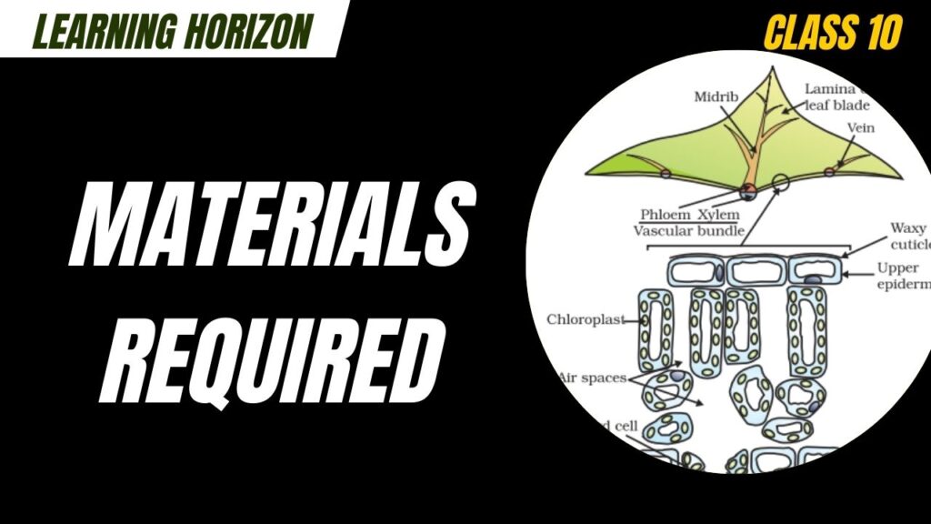
- Fresh dicot leaf (e.g., sunflower or hibiscus)
- Blade or razor
- Watch glass
- Safranin or iodine solution (staining agent)
- Microscope slides and coverslips
- Glycerine
- Light microscope
- Forceps
- Dropper
- Brush
Procedure:
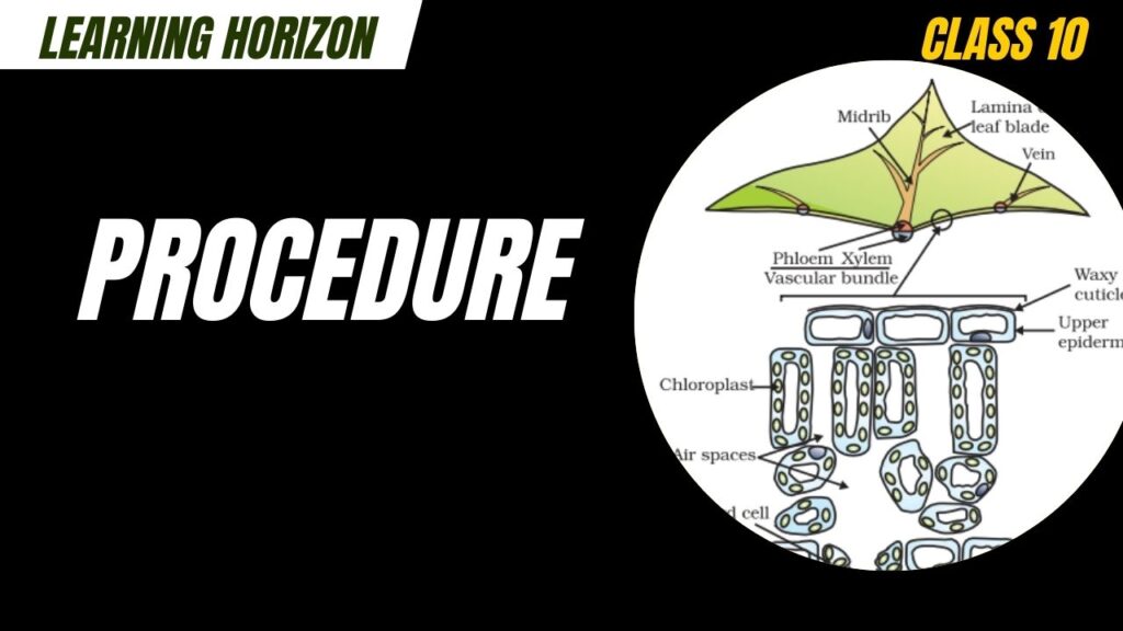
- Preparation of Leaf Section:
- Take a fresh dicot leaf.
- Using a blade or razor, carefully cut a thin transverse section (T.S.) of the leaf.
- The section should be thin enough to allow light to pass through when viewed under a microscope.
- Staining:
- Place the section in a watch glass containing safranin for about 2–3 minutes to stain the tissues.
- Carefully transfer the stained section to a slide using a brush.
- Mounting:
- Add a drop of glycerine on the section.
- Cover it with a coverslip gently to avoid air bubbles.
- Observation:
- Place the slide under the light microscope.
- Observe under low and high power to see different tissues.
Observations:
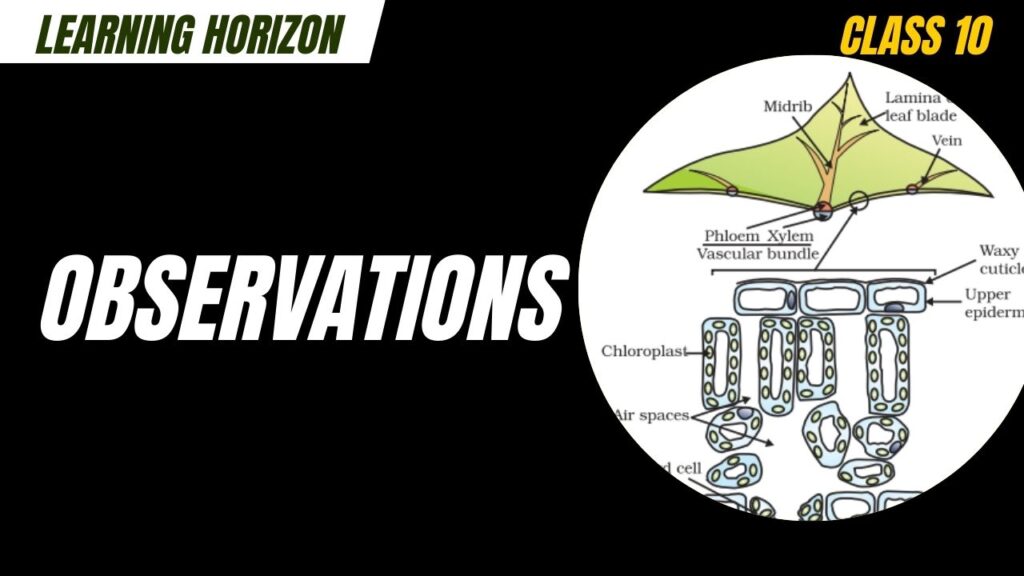
You will observe the following layers from upper to lower side:
- Upper Epidermis – A single layer of cells, often covered by a cuticle.
- Palisade Parenchyma – Tightly packed columnar cells rich in chloroplasts; major site of photosynthesis.
- Spongy Parenchyma – Loosely arranged cells with air spaces; also contains chloroplasts.
- Vascular Bundles (Veins) – Includes xylem and phloem; xylem transports water, and phloem transports food.
- Lower Epidermis – Contains stomata (pores) surrounded by guard cells for gas exchange.
Conclusion:
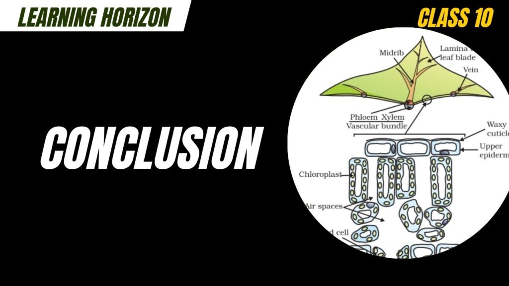
- The palisade mesophyll is the primary site of photosynthesis due to high chloroplast density.
- The vascular bundles play a key role in transport of water and food.
- The structure of the leaf is highly adapted to its function in photosynthesis and gas exchange.
1. What is Activity 5.1 in Class 10 Science?
Activity 5.1 involves observing the cross-section of a leaf under a microscope to study its internal structure and understand how leaves carry out photosynthesis.
2. What is the main aim of Activity 5.1?
The aim is to identify different parts of a leaf, such as epidermis, mesophyll, stomata, and vascular bundles, and understand their role in photosynthesis and gas exchange.
3. Which type of leaf is used in this activity?
A dicot leaf (like hibiscus or mustard) is used because it has clearly distinguishable tissue layers.
4. What materials are required for Activity 5.1?
- Fresh dicot leaf
- Blade or razor
- Staining solution (like safranin)
- Microscope slide and coverslip
- Glycerine
- Light microscope
5. Why is a stain like safranin used in this activity?
Stains like safranin help highlight plant cell structures, making it easier to see different tissues under a microscope.
6. What are the key parts seen in a leaf cross-section?
- Upper epidermis (outer protective layer)
- Palisade layer (rich in chloroplasts; main site of photosynthesis)
- Spongy layer (air spaces for gas exchange)
- Vascular bundles (xylem and phloem for transport)
- Lower epidermis with stomata
7. What is the function of stomata in the leaf?
Stomata are small pores that allow carbon dioxide to enter and oxygen to exit during photosynthesis. They also help in transpiration.
8. Why is it important to cut a very thin section of the leaf?
A thin section lets light pass through for clear visibility under the microscope and helps observe internal structures accurately.
9. What is observed in the palisade tissue?
The palisade cells are tightly packed and contain many chloroplasts, which are essential for capturing sunlight for photosynthesis.
10. What does Activity 5.1 teach Class 10 students?
It helps students visually understand how a leaf is structured to carry out photosynthesis efficiently and how different tissues play specific roles.